3D mammography technology is still considered relatively new in the health technology market, 2D mammography suffers from decreasing sensitivity as tissue density increases. This decreasing sensitivity can result in unclear images, which in turn may lead to cancers being missed, especially in patients with denser breasts.
Mammograms always present challenges to doctors in image reading and interpretation. A 3D object like the breast displayed as a 2D X-ray image effectively leads to a loss of image depth information. Lesions may be masked, due to image superimposition, by the tissue above or underneath, or normal structures may mimic a lesion.
Conventional analog mammography and full-field digital mammography display only the 3-D structure of the breast on a 2-D level, hampering the physicians’ efforts to identify certain types of tumours since anatomical structures in the breast can overlap and obscure lesions. Tomosynthesis acquires several breast projections from different angles and uses raw data to generate a 3-D volume dataset. Using this data set, clinicians can better analyze the type and size of breast lesions as well as microcalcifications compared to other forms of mammography. Breast tomosynthesis increases mammography’s sensitivity and specificity, in addition to improving efforts to differentiate and classify breast tumours.

The Digital Breast Tomosynthesis takes a series of about 15 to 50 images in an arc and then reconstructs them so it is similar to the doctor flipping through the pages of a book. It enables the doctor to detect cancer earlier than conventional 2D imaging, with the 3D image having higher clarity and definition.
Tomosynthesis, also known as digital tomosynthesis, is a method for performing high-resolution limited-angle tomography at radiographic dose levels. It has been studied for a variety of clinical applications, including vascular imaging, dental imaging, orthopaedic imaging, mammographic imaging, musculoskeletal imaging, and chest imaging.
The methods for reconstructing an arbitrary number of planes from a set of projections (the concept of tomosynthesis was derived and then developed from the work of Ziedses des Plantes.
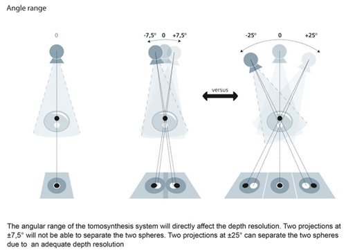
Digital tomosynthesis combines digital image capture and processing with simple tube/detector motion as used in conventional computed tomography (CT). However, although there are some similarities to CT, it is a separate technique. In CT, the source/detector makes a minimum 180-degree rotation around the patient obtaining a complete set of data from which images are computed. On the other hand Digital tomosynthesis only uses a limited rotation of 15-60 degrees with a lower number of discrete exposures (e.g., 7-51). This set of projections is digitally processed to yield images similar to conventional tomography with a limited depth of field. As the image processing is digital, a series of slices at different depths and with different thicknesses can be reconstructed from the same acquisition. However, since fewer projections are needed than CT to perform the reconstruction, radiation exposure and cost are both reduced.
Reconstruction algorithms for tomosynthesis are different from those of conventional CT because the conventional filtered back projection algorithm requires a complete set of data. Iterative algorithms based upon expectation maximisation are most commonly used, but are computationally intensive. Some manufacturers have produced practical systems using off-the-shelf processing units to perform the reconstruction in a few seconds.

Digital breast tomosynthesis (DBT) can provide a higher diagnostic accuracy compared to conventional mammography. In DBT, like conventional mammography, compression is used to improve image quality and decreases radiation dose.
Because the data acquired are very high resolution (85 - 160 micron typical), much higher than CT, DBT is unable to offer the narrow slice widths that CT offers (typically 1-1.5 mm). However, the higher resolution detectors permit very high in-plane resolution, even if the Z-axis resolution is less. DBT, as an extension to mammography, offers better detection rates with little extra increase in radiation.
Lesion visualization is superior with Digital breast tomosynthesis (DTM) when compared with digital mammography (DM), particularly of spiculated tumours. Although digital mammography (DM) is still the standard technique for imaging examination of symptomatic females, as well as for screening, it is a well-established fact that the technique has important limitations in terms of breast cancer detection, especially in dense breasts, where the sensitivity has been reported as being as low as 30–60%.
Sources:
http://www.live5news.com/story/30697712/3d-mammography-is-like-flipping-through-a-book
http://www.healthcare.siemens.com/mammography/be-sure/better-detection
https://en.wikipedia.org/wiki/Tomosynthesis
https://www.itnonline.com/content/fda-clears-siemens-breast-tomosynthesis-mammography-system
https://www.ncbi.nlm.nih.gov/pmc/articles/PMC4112403/
Compiled and edited by John Sandham.