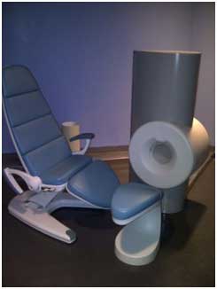
 Extremity MRI (Magnetic Resonance Imaging) scanners are technology specifically designed for the hands, wrists, elbows, feet, ankles and knees. Using the same technology as a full-body MRI scanner, a strong magnetic field and radio waves create high quality, detailed images of tissues and internal structures in these areas.
These images are fed back to a computer screen for a consultant to analyse. In some cases, extremity MRIs are able to depict issues such as fractures that may not show up on a standard X-ray. The extremity MRI scanner is more compact than a standard MRI because it only scans a limb at a time, not the whole body. This makes it a more comfortable experience for patients Extremity MRIs are painless, safe and don't involve radiation.
An extremity MRI smaller scanner is designed specifically for the body's extremities which eliminate the potential for claustrophobia, which some patients experience when enclosed in a full-body MRI machine. A traditional MRI requires you to lie completely still, but an extremity MRI won't limit your body movements quite as much.
One may undergo an extremity MRI to diagnose any of the following conditions in the arms, legs, hands, and feet:
Arthritis
Fractures
Bone infections
Tumours of the bone or soft tissue
Nerve-related issues
Stress injuries or injuries related to torsion or heavy impact
If an organisation has decided to get an Extremity MRI, there are 3 general types available:
High-field superconductive
Low-field permanent
Low-field permanent with limited shoulder capability
Pros for all types:
• They all have small footprints
• They will all give you good images for extremity scanning
• They will all cost less than a new standard MRI
Cons for all types:
• They only scan extremities
• You cannot image limbs that are larger than the bore (i.e. the limbs of severely obese patients)
• Now that you have a general picture of extremity MRI, let's break it down further and take a look at each type on its own.
High-Field Superconductive:
Example: ONI 1.0T Extremity MRI These come in 1.0T and 1.5T flavours. Both are hard to get on the secondary market, but 1.5T versions are especially rare at this point. The decision on 1.0 vs. 1.5 may simply come down to availability, but both approach the image quality level of standard magnets.
Pros:
• The best images available for extremity magnets
• No significant drop off of image quality from your standard full size MRI
• Unlikely to be removed from insurance reimbursement lists anytime soon.
Cons:
• Cannot image shoulders
• Uses cryogens (small amounts compared to standard magnets) so service costs are greater
Low-Field Permanent:
Example: Esaote 0.2T C-Scan Extremity MRI A scan on a 0.2T magnet will not replace primary MRI scans, but the low cost and small footprint of these magnets make it a way to free up some of your site's primary MRI capacity enabling a higher volume of larger examination scans, while still serving patients that only need an arm or leg scan.
Pros:
• Lowest cost
Cons:
• Cannot image shoulders
• .2T is a significant drop-off in image quality from standard full-size MRI
• Some insurers will not reimburse for scanning on a .2
Low-Field Permanent with Limited Shoulder Capability:
Example: Esaote 0.2T E-Scan XQ Extremity MRI
 All the time and space-saving benefits of low-field permanent magnets, plus some of the shoulder scanning capabilities of standard magnets.
Pros:
• Can image shoulders
• Relatively low price point
Cons:
• Similar image quality drop-off to the C-Scan
• Similar insurance concerns to the C-Scan
The benefits of an extremity MRI are ease of installation, ease of use, low maintenance, low energy consumption, no cryogens, and remote service, and reducing workload on the main MRI (for larger sites).
Extremity MRI systems use a fraction of the power employed by traditional MRIs saving thousands of £/Euro per year, due to the low power consumption (less than 3Kw in normal 220/110V power outlet); Low running costs; Cost effective: low break-even point.
Extremity MRI design integrates a complete MRI system including RF shielding, into one package, minimising the total space needed for installation. Due to the low weight and small magnet footprint, EMRI can be installed into much smaller facilities at much lower installation costs. The operator console can be located either inside or outside the scanning room, to accommodate both small diagnostic practices and large radiology departments. They are very compact and light-weight in comparison with a full MRIO facility. (Installation space of 9 m² (100 Sq Ft) required) and no RF shielding cage required. It can fits through standard clinic/hospital doorways.
Sources:
https://www.hcahealthcare.co.uk/about-hca/technology-at-hca/extremity-mri-scanner/
https://4rai.com/blog/3-types-of-mri-machines
https://info.blockimaging.com/bid/103103/Which-Extremity-MRI-Is-Right-for-Me
Compiled and edited by John Sandham


