
Positron Emission Tomography, or PET scanning, is a way of making images that show how the body works. In PET, a very small amount of a radioactive drug is given to a patient usually through an injection in the arm. The radioactive drug will go to certain parts of the body depending on which particular drug is used. For example, fluorine-18 labelled fluorodeoxyglucose, also called FDG, is a radioactive version of the sugar glucose (the isotope of fluorine is tagged to glucose, which has a half-life of about 112 minutes). Once injected into the body, it will go to places where glucose is used for energy. For instance, the brain uses glucose as its primary source of energy, and so FDG will go to the brain, and, in particular, to those parts of the brain that are actively burning glucose for energy. If the patient is performing a certain task, such as reading a story, more FDG will go to certain regions of the brain than others.
After being injected with the radioactive drug, the patient is placed in a machine called a PET scanner. The PET scanner consists of thousands of small radiation detectors that measure the radiation that is being emitted by the radioactive drug within the patient. In this way, the PET scanner can make 3D images that show where the radioactive drug went in the body. In the case of FDG, these images will show those areas of the body that are actively burning glucose for energy. A PET scan shows how the body is working and not just how it looks as other forms of medical imaging, such as CAT scanning and MRI, do. Other radioactive drugs besides FDG can also be used to measure other aspects of how the body works such as the rate of blood flow in certain organs and how certain cells in the body, such as nerve cells, communicate with each other.
In many cases, PET scanning is used to either find a disease or to better understand how widespread a disease is within a patient. PET scanning is commonly used for such diseases as epilepsy, Alzheimer’s disease and cancer. In epilepsy, it can be used to tell where within the brain the disease is and whether the patient is a candidate for certain methods of treating the disease. In Alzheimer’s disease, the PET scan can be used to tell if this patient truly has the disease or whether the patient’s symptoms (such as forgetfulness) are actually being caused by something else.
In cancer, PET scanning can be used to find a tumour. It can also be used to tell whether a tumour found by some other method is cancerous or whether the cancer has spread to other parts of the body. PET scanning can do this because FDG is known to go to many types of cancerous tumours at a much higher rate than normal tissue.
PET scanning is also used in research to gain a better understanding of how our bodies work, learning about Alzheimer’s disease, dyslexia and learning disabilities, drug abuse, and cancer, as well as the biological effects of social stress and normal ageing.
 PET scan of Lung Tumour
PET scanning is addressing many important issues in medicine as well as in basic biology. In the years to come, as we learn more about the fundamental nature of many diseases, PET scanning will be an incredibly invaluable tool as it continues to provide a unique opportunity to look inside and see how the body works.
Unlike other types of scans, such as computerised tomography (CT) and MRI, PET scans distinguish whether a tumour is malignant as well as monitor the progress of cancer treatments. The implications for cancer treatment centres are enormous.
While some argue that PET scans could replace a number of other more traditional scans done in radiology, that does not appear to be the case-as of yet. Most physicians and scientists working with this new technology believe that PET is a complimentary test that works in concert with the other tests and can provide information that may not be evident on the other scans.
Patients who undergo a PET scan for cancer diagnosis receive an injection of fluorodeoxyglucose FDG. After about 30 to 45 minutes when the body absorbs the FDG, patients lie down while they move through the PET scanner.
Cancerous cells absorb more of the radioactive glucose than normal cells since cancer cells metabolise glucose at a rate of approximately 20 times that of normal tissue. As the tumour absorbs glucose, the fluorine goes with it and emits a signal that is detected by the PET camera. The PET scanner then assembles these high-energy particles into an image. Areas of concern are lighted up in a three-dimensional image for physicians to review.
PET differs from MRI in that it can create images of metabolic activity. Combining that with anatomical images from MRI gives doctors a moving picture of what's happening inside the body, with information from the molecular level.
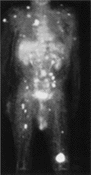 PET Full body image
As an example of the power of this technology, consider a patient with a tumour indicated in one location, with CT images and biopsies to back it up. Initially, the patient is regarded as a candidate for surgery. However, the PET shows diffusion of cancer cells with such clear and detailed images that the doctors can make a treatment decision to redirect the patient to chemotherapy.
PET scans may be useful in modifying treatment regimes since the PET scan can determine if a tumour has been completely eradicated or if the tumour is partly active. In the former situation, a patient undergoing a particularly rigorous regime may be able to cease treatment. In the latter situation, additional or adjunctive treatment may be recommended.
By detecting molecular activity, doctors also can use a PET scan for post-treatment follow up checks. While a CT image will show a mass of tissue, a PET scan can determine if this mass is leftover scar tissue or a recurring, active tumour.
Isotopes
Isotopes are the keys to timely patient treatment. Generally, isotopes are produced by huge, very expensive cyclotron devices. A cyclotron is a particle accelerator which produces positron-emitting elements or short-lived radioisotopes. These radioisotopes can then be incorporated into other chemical compounds which are synthesised into a final product that can be injected into a person. These radioisotopes are used to "label" compounds so it can later be identified where in the body the radiopharmaceutical is being distributed. The compounds that are being labelled are organic molecules normally utilised in the body such as sugar, neurotransmitters, etc...
First, the cyclotron bombards non-radioactive elements in the target with accelerated particles which converts these elements into positron-emitting radioactive isotopes of fluorine, nitrogen, oxygen, or carbon. The current radioactive isotope produced at our site is fluorine-18 (F-18) which has a half-life of 110 minutes. F-18 thus produced from the cyclotron is delivered to a chemical synthesis unit called the chemical processing unit (CPCU). This is where F-18 is incorporated into a precursor to produce the final product, the labelled sugar molecule fluorodeoxyglucose (FDG). This entire process is fully automated and done in the cyclotron lab. When a dose is needed, it is transported to the PET scan room by way of a dedicated pneumatic tube system.
In fact, one of the widespread barriers to PET use has been the need for a cyclotron, which costs more than the scanner itself, and requires a full-time physicist to operate.
Cardiology has had a longer history with PET scanners. In cardiology, they use an isotope of rubidium, which releases the positron. Cardiologists don't need access to a cyclotron because rubidium 82(82Rb) can be created through a chemical reaction in a generator. In cardiology, PET scanners answer the question of whether the heart is viable after a heart attack.
Another isotope, radioactive oxygen, provides the first reliable diagnosis of Alzheimer's disease in the living. (Otherwise, it can only be confirmed post-mortem).
A fixed PET scan unit can be placed into an existing diagnostic or treatment space with a certain amount of renovation or an addition may be built to house the PET scanner as well as required support spaces. Approximately 2,000 square feet are needed to house a full PET scan operation although this can change substantially depending upon individual circumstances. Typically included are the PET scan room that resembles a CT scan facility, a hot lab (if not shared), equipment control room, patient waiting, patient prep area, and a patient toilet. Some spaces may need to be lead lined, depending on uses of adjacent space. Patient "throughput" ranges from 45 minutes to one hour and 15 minutes to complete a scan, depending on equipment.