
There have been truly incredible strides in the standard of burn care. The mortality from burn injuries has more than halved since the 1950s, making it hugely unique among major diseases of the developed world. There can be no doubt technology and technological advances have driven this process, dramatically improved every aspect of burn care, from the intensive care management, the surgical management, management of the healing wound to the post burn sequelae, specifically scar management. This review aims to identify key technological advances in burns, in both the developed and developing world, and evaluate their influence in the continued strategy to improve the standards of global burn care.
There are three levels of burns:
- First-degree burns affect only the outer layer of the skin. They cause pain, redness, and swelling.
- Second-degree (partial thickness) burns affect both the outer and underlying layer of skin. They cause pain, redness, swelling, and blistering.
- Third-degree (full thickness) burns extend into deeper tissues. They cause white or blackened, charred skin that may be numb.
Technology for burn victims
Specialized medical equipment is crucial to the recovery of burn victims: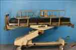
- Electric Beds: - Patients can manipulate their own bed position and are more independent.
- Specialist Microscopes: - Plastic surgeons can perform micro-vascular surgery.
- Mobilise Stretcher: - Easy transfer of patient to and from bed with minimal patient movement and a built-in scale for weighing and immobilized patient.
- Patient Controlled Analgesic Pumps: - Patients can manage their own pain by pushing a button which releases medication.
Artificial skin
Burned, dead skin must be replaced with something that does everything the old skin did: regulate temperature, keep in fluids and keep out invaders like bacteria.One option is to move a thin layer of healthy skin from elsewhere on the body to cover the burn. But if more than 50 percent of the body is burned, there obviously isn't enough healthy skin. Skin from cadavers can help temporarily, but after a few weeks will be rejected by the body's immune system.
Artificial skin nurtures the body's own skin cells, sometimes even deceiving them as they struggle to grow and replace burned tissue. The technology is promising, but doctors warn it's costly and not always completely effective.Spray-on cells to treat severe burns
A team at Queen Victoria Hospital in East Grinstead, West Sussex, has used the technique to treat several patients - including a man with 90% burns.The latest study examines whether the cells go on to become a fully functioning part of the skin. The technique was first pioneered in Perth, Western Australia, and used on some patients - including victims of the Bali bombing - but has never been fully evaluated. It can cover a much bigger areas and do it much more quickly. The East Grinstead team have embraced the technology, and used it to treat several patients with severe and extensive burns.
Mesher
The current method of treating burns victims is to take samples of skin from unaffected areas, and put them through a meshing machine. 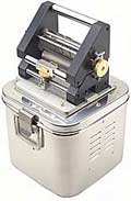
The mesher's platform will accommodate almost any size and irregular shape of skin graft. No lubrication is required and the design allows easy access for cleaning, which makes it virtually maintenance free and autoclavable.
This expands the tissue, creating a string vest pattern of connected patches of skin surrounded by large holes.
Slow method: The technique can be used to cover big patches of tissue where the skin has been completely burned away. However, it is slow, and not always effective.
New technique
The East Grinstead team are using a new technique that has the potential to be much more useful. The treatment helped heal severe burns on an elderly patient's legs.
Mr Phil Gilbert, a consultant plastic surgeon who specialises in burns said: "It can cover a much bigger areas and do it much more quickly."In pilot studies we also get the impression that wounds heal noticeably quicker with less scarring."
A healthy skin sample is taken from the patient, and split in the laboratory to separate out the surface cells, known as keratinocytes. These cells are then cultured for two to three weeks, and made up into a suspension.
At the same time other skin cell tissue from the patient is put through a different type of meshing machine, known as a meek mesher. Instead of creating a string vest pattern of tissue, this machine cuts the skin sample into tiny little squares.Tissue combination
The cultured cells are then sprayed on to the small pieces of tissue and combine to create new skin for the patient. Mr Gilbert said the technique had been used to treat a man who suffered 90% burns. Initially his burns were shaved off, and he was covered in sterile skin which had been harvested from bodies and kept frozen in storage. Doctors were only able to take skin samples for culture from small areas of one leg unaffected by burns. "We would have struggled to keep him alive using the standard methods," he said. "These have been used to treat people with very extensive burns, but these have nearly always been children." His skin is far from normal - it is thin, and has no hair follicles, sweat or oil glands. But it has begun to settle down, and take on a more natural pale colour.Strict regulations on the storage of skin, introduced in the wake of concerns over CJD, mean that it is very expensive to culture cells in this way. "People want to know why they should spend x thousand pounds on a patient, so the stronger the evidence we can produce that is worthwhile the better," he said.
Scar management
Successful approaches to modulate the scar remodeling process, minimise the development of scarring, specifically hypertrophic scarring, have yet to be established. Proposed beneficial effects of novel surgical techniques are awaited. Meanwhile scar management remains a formidable challenge for the burn community. Currently the most commonly used techniques are pressure garments, with and without silicone and injected corticosteroids. Evidence has been conflicting with studies showing modest to no improvements with use of these techniques. Numerous novel therapies have been introduced, but to date, have not made the anticipated impacts. The future of wound healing and scar modulation is thought to hinge on our growing understanding of progenitor and stem cells and from development of these novel therapies.
Laser doppler imaging
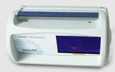 Dozens of children in the UK receive a serious burn or scald every day and are rushed to hospital emergency departments, where burns specialists assess their injuries and decide on treatment, which may include skin grafts and surgery. Laser doppler imaging (LDI) is a vital tool in this assessment. By measuring blood flow beneath the burn wound, LDI allows doctors to accurately assess the extent of the injury. Deeper and severe burns require skin grafts and surgery, which may lead to scarring and prolonged and painful recovery, whereas less severe cases can heal without such intervention.
Medical technology pioneers at the University of Nottingham have equipped doctors across the world with smaller, faster LDI scanners, which help ease the suffering of thousands of young burns victims every year. They are scanned at the bedside in seconds, minimising the need for additional sedation or prolonged exposure of the wound. The devices are saving the NHS and health providers across the world millions of pounds by cutting the cost of repeated scans, and many millions more by helping to identify cases where surgery is not necessary, which in turn reduces hospital stays and shortens rehabilitation.
Professor Steve Morgan, of the Optics and Photonics Group (OPG) at Nottingham,who led the research alongside Professor Barrie Hayes-Gill, Professor John Crowe and Dr Yiqun Zhu, said: "LDI has been recognised as a key tool in burns assessment for a number of years and the UK’s Moor Instruments is a global leader in the field. However, LDI typically uses single-point imaging and requires the patient to remain still for three minutes to get a clear image. That’s a big task for any burns victim, especially for a small child in distress. Moor approached us to help develop a high-speed scanning device and the project won funding from Innovate UK”.
“We’ve worked with Moor to replace single-point imaging with an array of laser lights scanned across the tissue surface. Our advances in sensing and the processing of an array of signals, and in developing new, faster and more dynamic camera chips, means we can capture an image in 3 to 4 seconds.” An independent clinical trial found the Moor LDLS-BI device had a 94.6% accuracy for predicting the potential for healing of burn wounds by allowing doctors to ‘see’ beyond the skin surface. Andrew Holland, Professor of Paediatric Surgery at The University of Sydney School of Medicine and the Children’s Hospital at Westmead Clinical School, Australia, said: “We use the moorLDLS for paediatric patients approximately 10 times every month. We have found the scanners very useful in prioritising patients for surgery and planning their elective procedures. I am committed to improving the care of burn and other traumatic injuries in children and the moorLDLS has been crucial in helping me deliver on this promise.” This lightweight, portable device is now commercially available and is used every day in paediatric burns units, easing suffering and improving outcomes for about 5,000 children a year. The rapid scanning of the moorLDLS has made LDI scanning more accessible to children. This imaging supports better decision-making about skin grafting and is estimated to save healthcare providers globally around £6m per year.
Professor Morgan added: “It’s immensely gratifying that our collaboration with Moor Instruments has resulted in these scanners being used in burns units in the UK and across the world.” Dr Rodney Gush of Moor Instruments said: “The University of Nottingham is a world-leader in translating sensing and imaging research into clinical practice. Our partnership underlines that continual innovation lies at the heart of advances in healthcare.”
Sources:
http://news.bbc.co.uk/2/hi/health/4208746.stm
https://www.templehealth.org/about/blog/spray-skin-technology-heal-severe-burns#:~:text=Rae%3A%20Yes%2C%20the%20FDA%20recently,few%20hospitals%20that%20offers%20it
https://www.ncbi.nlm.nih.gov/pmc/articles/PMC6146166/
https://www.nottingham.ac.uk/vision/transforming-outcomes-for-young-burns-victims
https://www.moor.co.uk/products/previous-system/moorldi/
