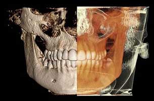
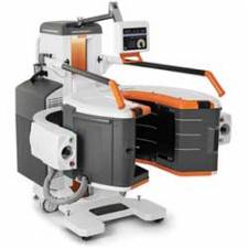
 Cone beam computed tomography is a special type of x-ray equipment used when regular dental or facial x-rays are not sufficient.
Your doctor may use this technology to produce three dimensional (3-D) images of your teeth, soft tissues, nerve pathways and bone in a single scan.
This type of CT scanner uses a special type of technology to generate three dimensional (3-D) images of dental structures, soft tissues, nerve paths and bone in the craniofacial region in a single scan. Images obtained with cone beam CT allow for more precise treatment planning.
Cone beam CT is not the same as conventional CT.
However, dental cone beam CT can be used to produce images that are similar to those produced by conventional CT imaging. Cone beam computed tomography (or CBCT, also referred to as C-arm CT, cone beam volume CT, or flat panel CT) is a medical imaging technique consisting of X-ray computed tomography where the X-rays are divergent, forming a cone.
Cone beam technology was first introduced in the European market in 1996 by QR s.r.l. (NewTom 9000) and into the US market in 2001. October 25, 2013, during the "Festival della Scienza" in Genova, Italy, the original members of the research group: Attilio Tacconi, Piero Mozzo, Daniele Godi and Giordano Ronca received an award for the cone-beam CT invention, a revolutionary invention that changed world's dental radiology panorama.
During a cone beam CT examination, the C-arm or gantry rotates around the head in a complete 360-degree rotation while capturing multiple images from different angles that are reconstructed to create a single 3-D image.
The x-ray source and detector are mounted on opposite sides of the revolving C-arm or gantry and rotate in unison. In a single rotation, the detector can generate anywhere between 150 to 200 high resolution two-dimensional (2-D) images, which are then digitally combined to form a 3-D image that can provide your dentist or oral surgeon with valuable information about your oral and craniofacial health.
 Some manufacturers are able to combine three different types of 3D data with one X-ray unit, bringing together a Cone Beam Computed Tomography (CBCT) image; 3D face photo and 3D model scan into one 3D image.
This 3D combination creates a virtual patient in 3D, helping consultants with their clinical practice.
 CBCT has become increasingly important in treatment planning and diagnosis in implant dentistry, ENT, orthopaedics, and interventional radiology (IR), among other things.
Perhaps because of the increased access to such technology, CBCT scanners are now finding many uses in dentistry, such as in the fields of oral surgery, endodontics and orthodontics. Integrated CBCT is also an important tool for patient positioning and verification in image-guided radiation therapy (IGRT).
During dental/orthopaedic imaging, the CBCT scanner rotates around the patient's head, obtaining up to nearly 600 distinct images. For interventional radiology, the patient is positioned offset to the table so that the region of interest is centred in the field of view for the cone beam. A single 200 degree rotation over the region of interest acquires a volumetric data set. The scanning software collects the data and reconstructs it, producing what is termed a digital volume composed of three-dimensional voxels of anatomical data that can then be manipulated and visualized with specialized software.
The patient will be asked to remain very still while the x-ray source and detector revolve around their head for a 360-degree rotation or less. This typically can take between 20 to 40 seconds for a complete volume, also called a full mouth x-ray, in which the entire mouth and dental structures are imaged, and less than 10 seconds for a regional scan that focuses on a specific area of the maxilla or mandible.
Dental cone beam CT is commonly used for treatment planning of orthodontic issues. It is also useful for more complex cases that involve:
• Surgical planning for impacted teeth.
• Diagnosing temporomandibular joint disorder (TMJ).
• Accurate placement of dental implants.
• Evaluation of the jaw, sinuses, nerve canals and nasal cavity.
• Detecting, measuring and treating jaw tumours.
• Determining bone structure and tooth orientation.
• Locating the origin of pain or pathology.
• Cephalometric analysis.
• Reconstructive surgery.
Benefits
• The focused x-ray beam reduces scatter radiation, resulting in better image quality.
• A single scan produces a wide variety of views and angles that can be manipulated to provide a more complete evaluation.
• Cone beam CT scans provide more information that conventional dental x-ray, allowing for more precise treatment planning.
• CT scanning is painless, non-invasive and accurate.
• A major advantage of CT is its ability to image bone and soft tissue at the same time.
• No radiation remains in a patient's body after a CT examination.
• X-rays used in CT scans should have no immediate side effects.
Risks
There is always a slight chance of cancer from excessive exposure to radiation. However, the benefit of an accurate diagnosis far outweighs the risk.
Because children are more sensitive to radiation, they should have a CT exam only if it is essential for making a diagnosis and should not have repeated CT exams unless absolutely necessary. CT scans in children should always be done with low-dose technique.
Sources:
https://www.radiologyinfo.org/en/info.cfm?pg=dentalconect
https://en.wikipedia.org/wiki/Cone_beam_computed_tomography
http://www.planmeca.com/Imaging/3D-imaging/
Edited by John Sandham.


