
 Medical Thermography (digital infrared thermal imaging - DITI ) is used as a method of research for early pre-clinical diagnosis and control during treatment of homeostatic imbalances.
There are few devices, which operate in a passive method like infrared Thermography medicine; amongst these are the ECG and EEG. The intrinsic safety of this method makes infrared Thermography free from any limitations or contra- indications.
Thermography is a non-invasive, non-contact tool that uses the heat from your body to aid in making diagnosis of a host of health care conditions. Thermography is completely safe and uses no radiation.
Medical Thermography equipment usually has two parts, the IR camera and a standard PC or laptop computer. These systems have only a few controls and relatively easy to use.
Monitors are high-resolution full colour, isotherm or grey scale, and usually include image manipulation, isothermal temperature mapping, and point-by-point temperature measurement with a cursor or statistical region of interest. The systems measure temperatures ranging from 10° C - 55° C to an accuracy of 0.1° C. Focus adjustment should cover small areas down to 75 x 75mm.
These systems are PC based and therefore able to store tens of thousands of images (and these images may be retrieved for later analysis). The ability to statistically analyse the thermograms at a later date is very important in clinical work. Copies of images can easily be sent (via e-mail, floppy disk, etc.) to referring doctors or other healthcare professionals.
The medical applications of DITI are extensive, particularly in the fields of Rheumatology, Neurology, Oncology, Physiotherapy and sports medicine. Thermal imaging systems are an economical easy-to-use tool for examining and monitoring patients quickly and accurately.
Utilising high-speed computers and very accurate thermal imaging cameras, the heat from your body is processed and recorded in the computer into an image map which can then be analyzed on screen, printed or sent via email.
A doctor can then use the image map to determine if abnormal hot or cold areas are present. These hot and cold areas, can relate to a number of conditions for which the Food and Drug Administration, Bureau of Medical Devices has approved the thermography procedure. These include, the screening for breast cancer, extra-cranial vessel disease (head and neck vessels), neuro-musculo-skeletal disorders and vascular disease of the lower extremities.
There have been a number of advancements in the past decade, which has brought thermal imaging in medicine back to the forefront of diagnosis. As technology has advanced, so has our "medical" concept of thermal imaging.
Some of the common applications of Thermography are in:
- Breast pathologies
- Extra-Cranial Vessel Disease
- Neuro-Musculo-Skeletal
- Vertebrae (nerve problems/arthritis)
- Lower Extremity Vessel Disease
Breasts pathologies
Probably the most applied area of Medical Thermography - breast cancer, benign tumours, mastitis, and fibrocystic breast disease.
The utilization of thermography as a screening tool in the detection of breast cancer has been for the past decade a very controversial topic within the health care community.
However, the technology has gained in scientific acceptance, has been approved for screening purposes and is clearly a powerful tool in the war on breast cancer.
The concept is quite simple. Thermography measures the heat coming from your body. Metastatic cancers create heat which can be imaged by digital infrared imaging. This is due to two separate yet connected factors.
The first is the metabolic activity of the tumour tissue as compared with the temperature of tissue adjacent to the tumour, and in the opposite breast. By comparing the breast in question with the normal breast which acts as the patients own control, abnormal heat signatures associated with the metabolism of the tumour can be detected easily. These differences in temperature are referred to as a Delta T.
The second method of detection is due to the angiogenesis of the tumour.
i.e. Cancerous tumours produce a chemical which actually promotes the development of blood vessels supplying the area where the tumour resides. Also, normal blood vessels which are under the control of the sympathetic nervous system are essentially paralyzed, causing vaso-dilation, or an increase in size of the blood vessel. The increase in blood in the region due to angiogenesis and combined with the vaso-dilation simply means more heat, recordable with thermal imaging procedures.
As thermal imaging has been demonstrated in numerous studies to be capable of measuring these heat signatures years before conventional technologies can see a mass, and as the procedure uses no radiation, compression of breast tissue and as it is totally safe, thermography or DITI provides for a safe early warning detection system.
Extra-Cranial Vessel Disease
In a similar way, a variety of conditions which relate to flow of blood through the vessels of the neck and head are readily accessed with thermal imaging. As the blood vessels in the face and skull are coursing through very thin tissue between the bones of the skull and the skin covering the skull, they are readily and easily visualized with thermal imaging.
As the vessels of the neck are very large calibre vessels, they too are very easily visualized with thermography and clues to the potential of developing vascular disease which might lead to stroke are a consideration when performing thermography.
The use of thermography in differentiation of various types of headache (migraine, cluster, cervical spine related), facial nerve injury as in the case of a blow to the face or a car accident where the face contacts a windshield or the steering wheel, the visualization of TMJ disorders (tempero-mandibular joint) are commonly used aspects of thermographic diagnosis and analysis of the head and neck.
The ability of thermal imaging to safely indicate the heat from sources in the jaw and teeth is providing a very exciting opportunity to screen individuals for dental decay and cavitations without routine screening x-rays. Also, a number of patients have been seen with heat signatures in the jaw related to amalgam fillings which might be toxic for that particular patient. This area of thermal imaging is very promising.
Neuro-Musculo-Skeletal
This is one of the clearest examples of thermography's ability to accurately diagnose patients with a host of back, neck and extremity disorders. In fact, it was the use of thermography by chiropractors, neurologists and orthopaedists in the late 70's to the late 80's in spinal injury cases from car accidents and work injuries, which really launched the clinical interest in this diagnostic tool.
When muscle tissue is strained or torn, it releases chemicals which cause increased heat. This can be seen as intense patterns of hyperthermia in the region of the muscle, or trigger point, as in the case of fibromyalgia. Heat patterns can also be seen in the legs and soles of the feet which indicate altered gait or weight bearing mechanics, which might relate to a low back or foot condition.
Further, back strain produces very consistent heat patterns which not only tell us about the source of probable spinal injuries, but can also tell us about areas of spinal compensation, In effect, a low back might be being treated by a chiropractor, when the mid back or neck is actually the source of the problem.
Nerve damage, as occurs in disc herniation and spinal nerve root compression displays on the thermographic map in exactly the opposite direction as muscle injury by revealing cool areas of hypothermia in the nerve tracts coming from the spine. In this way, thermography can demonstrate and document permanency of spinal injuries which are causing a person disability. This documentary, not diagnostic aspect of thermography has been used for many years in the trial courts to prove injury and assist in the rating of permanent impairment.
Lower Extremity Vessel Disease
The ability of thermography to detect the presence of deep vein thrombosis and other circulatory disorders of the lower extremities is a very exciting application of this procedure as it allows us to painlessly and safely detect possible disease that if unchecked, could cause the loss of a limb, or in some cases add to the possibility of stroke.
Another aspect of thermal imaging which has gone largely unnoticed is in developing diabetic neuropathies of the feet, before the foot becomes insensate.
For example we often see individuals who have extremely cold feet thermographically, although they have no other symptoms.
The feet demonstrate thermographically as 1-2 degrees centigrade colder than the lower leg, and usually the toes are not visible to the camera as they have become so hypothermic. This can occur several years before routine blood tests indicate diabetes, and as such, can give the patient time to treat the condition before permanent nerve damage occurs to the foot.
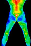
 These images are pre and post thermograms of a patient suffering from diagnosed fibromyalgia of many years duration.
The upper thermogram is prior to treatment. There are 4 primary sites of involvement (see explanation below) which are obvious on the upper thermogram.
Following treatment using spray and stretch techniques and specific chiropractic manipulation with deep tissue massage, she improved significantly.
Muscular hyperthermic patterns at the left of approximately the first cervical vertebra consistent with trauma and biomechanical instability (chronic) Trigger point irritation at the right levator scapulae muscle (chronic) consistent with a primary site.
Increased heat signature at the posterior SUPRASPINOUS LIGAMENT consistent with residual chronic irritation from a thoraco-lumbar sprain strain injury. Very focal hyperthermia at the midline of the lumbar spine approximating L1-L2 and consistent with biomechanical instability related to severe sprain strain and/ or lumbar disc involvement.
Other areas where medical Thermography is successfully applied:
Respiratory dysfunctions
Infrared Thermography was applied during the last epidemic of atypical pneumonia (SARS) at airports and is useful for monitoring asthma, allergies, bronchitis, influenza etc.
Digestive disorders
Infrared Thermography has demonstrated excellent results in helping in the diagnosis of urgent gastrointestinal pathology, especially appendicitis, irritable bowel syndrome (IBS), colitis, ulcerative colitis and hyper and hypo gastric secretions.
Urinary diseases
infrared Thermography helps to save patient's and doctor's time in waiting for laboratory data and is successfully used to monitor Urinary tract infections, kidney pathology etc.
Cardiovascular and circulatory disorders
Infrared Thermography is periodically applied for differential diagnostics and is useful in preventing heart disease and serious circulatory problems such as varicose veins. Specific valvular points can be located for surgical purposes as well as treatment suggestions.
Lymphatic dysfunctions
Infrared Thermography tests therapy effectiveness in severe cases of lymphoma, leukaemia and reliable to monitor lymphatic involvement in breast cancer patients.
Reproductive disorders
Infrared Thermography has its own specific application in gynaecological problems, uterus, prostate and polycystic ovaries, endometriosis and fibroids.
Nervous dysfunctions
Infrared Thermography analyses the brain, spinal cord and nerves, gives doctor a reliable and safe method of problem location and for monitoring improvements.
Endocrine Disorders
Infrared Thermography helps to evaluate hormonal changes, thyroid disorders such as hypo and hyperthyroidism, and diabetes
Locomotors Disorders
Infrared Thermography helps in the clinical evaluation and detection of serious and difficult disorders such as musculo-skeletal syndromes, neuropathy, neurovascular compression, nerve injury, soft tissue injury, arthritis, carpel tunnel syndrome, myofascial syndromes, inflammatory pain, and disk injury.
Surgical Assistance
Surgeries can be assisted safely before and after using Medical Thermography- helps to locate tumours size and locates surgical area and monitors the healing process after surgery.
Skin Problems
Infrared thermography gives a more precise level of information - skin tumours and skin cancers, and wound healing.
Ear, Nose, and Throat dysfunction
Infrared thermography can assist in identifying areas with disorders when radiation should not be used such as tonsillitis, swelling of the lymphatic glands, rhinitis, teething problems, sinusitis, and otitis.
Dentistry
Dentists recommend the use of Medical Thermography in monitoring control in the inflammation process into oral cavity and reaction of the regional lymphatic nodes, maxillary joint disease and other chronic diseases of the bones, nerves, located in the maxilla facial area. Medical Thermography can also measure temperature changes in the application of new methods and dental materials applied by dentists.
Sources:
http://66.241.252.6/thermography.html
http://www.icim.ie/medical-thermography.asp
http://www.fdlac.com
http://www.jkns.or.kr
http://www.meditherm.com/therm_default.htm


![Functional Magnetic Resonance Imaging [fMRI]](/images/thumbnails/mod_relateditems_xtd/36_199.jpg)

