 Vascular ultrasound provides pictures of the body's veins and arteries. Ultrasound imaging is based on the same principles involved in the sonar used by bats, ships, fishermen and the weather service. When a sound wave strikes an object, it bounces back, or echoes. By measuring these echo waves, it is possible to determine how far away the object is and its size, shape and consistency (whether the object is solid, filled with fluid, or both).
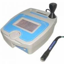
In medicine, ultrasound is used to detect changes in appearance of organs, tissues, and vessels or detect abnormal masses, such as tumors. In an ultrasound examination, a transducer both sends the sound waves and receives/records the echoing waves. When the transducer is pressed against the skin, it directs small pulses of inaudible, high-frequency sound waves into the body. As the sound waves bounce off of internal organs, fluids and tissues, the sensitive microphone in the transducer records tiny changes in the sound's pitch and direction. These signature waves are instantly measured and displayed by a computer, which in turn creates a real-time picture on the monitor. One or more frames of the moving pictures are typically captured as still images. Small loops of the moving "real time" images may also be saved.
Typical diagnostic sonographic scanners operate in the frequency range of 2 to 18 MHz, but frequencies from 50 to100 MHz have been used experimentally in a technique known as biomicroscopy in special regions, such as the anterior chamber of the eye. The choice of frequency is a trade-off between spatial resolution of the image and imaging depth: lower frequencies produce less resolution but image deeper into the body.
Higher frequency sound waves have a smaller wavelength and thus are capable of reflecting or scattering from smaller structures. Higher frequency sound waves also have a larger attenuation coefficient and thus are more readily absorbed in tissue, limiting the depth of penetration of the sound wave into the body.
 Sonographers typically use a hand-held probe (called a transducer) that is placed directly on and moved over the patient.
Seeing deep into the body with sonography is very difficult. Some acoustic energy is lost every time an echo is formed, but most of it (approximately 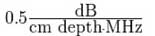 ) is lost from acoustic absorption.
Vascular ultrasound, a special application of ultrasound, measures the direction and speed of blood cells as they move through vessels. The movement of blood cells causes a change in pitch of the reflected sound waves (called the Doppler effect). A computer collects and processes the sounds and creates graphs or color pictures that represent the flow of blood through the blood vessels.
 Vascular ultrasound of the arteries, such as the carotid arteries supplying the brain with blood, can help in identifying areas of narrowing (stenosis), calcifications or thrombosis that may lead to a stroke. It will help your doctor in planning your treatment.
It can also identify areas of abnormal widening of blood vessels (aneurysm) that, if left untreated, can lead to serious consequences.
An example vascular ultrasound image is shown of the Carotid artery branching into external and internal divisions. Patient has head to left and feet to right.
Ultrasound of the veins can identify thrombosis (blockage) that may be the cause of leg or arm swelling or pain. In people with varicose veins, it can help to identify the source of the supply of these veins and help the surgeon decide how best to deal with this condition
Strengths
It renders "live" images, where the operator can dynamically select the most useful section for diagnosing and documenting changes, often enabling rapid diagnoses. Live images also allow for ultrasound-guided biopsies or injections, which can be cumbersome with other imaging modalities.
It shows the structure of organs.
It has no known long-term side effects and rarely causes any discomfort to the patient.
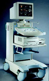
Equipment is widely available and comparatively flexible.
Small, easily carried scanners are available; examinations can be performed at the bedside.
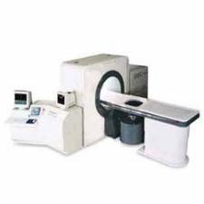
Relatively inexpensive compared to other modes of investigation, such as computed X-ray tomography, DEXA or magnetic resonance imaging.
Spatial resolution is better in high frequency ultrasound transducers than it is in most other imaging modalities.
Weaknesses
Sonographic devices have trouble penetrating bone. For example, sonography of the adult brain is very limited though improvements are being made in transcranial ultrasonography.
Sonography performs very poorly when there is a gas between the transducer and the organ of interest, due to the extreme differences in acoustic impedance. For example, lung imaging is not possible (apart from demarcating pleural effusions).
The depth penetration may be limited depending on the frequency of imaging. Consequently, there might be difficulties imaging structures deep in the body, especially in obese patients.
Body habitus has a large influence on image quality, image quality and accuracy of diagnosis is limited with obese patients, overlying subcutaneous fat attenuates the sound beam and a lower frequency transducer is required (with lower resolution).
The method is operator-dependent. A high level of skill and experience is needed to acquire good-quality images and make accurate diagnoses.
Sources:
http://www.radiologyinfo.org/en/photocat/gallery3.cfm?pid=1&image=carotid-Dus-brnch5.jpg&pg=vascularus
(Link no longer functional.)
http://www.kentmedicalimaging.co.uk/vascular.htm http://www.svtgbi.org.uk/
http://en.wikipedia.org/wiki/Medical_ultrasonography
Compiled and edited by John Sandham