An echocardiogram, often referred to in the medical community as a cardiac ECHO or simply an ECHO, is a sonogram of the heart. Also known as a cardiac ultrasound, it uses standard ultrasound techniques to image two-dimensional slices of the heart. The latest ultrasound systems now employ 3D real-time imaging.
What is Ultrasound?
Sound is made up of several different frequency waves. The very high frequency range is inaudible to the human ear and is known as ultrasound. Ultrasound was used by the Navy during World War II to detect submarines, and is widely used by fisherman to help find schools of fish.
 In each case, an ultrasound machine is used. With the help of a microphone-shaped device (known as a transducer) ultrasound waves are created and beamed through water. When the beam encounters a boundary or interface between liquid (water) and a solid (submarine or fish) with a different density or compactness, part of the beam is reflected back to the transducer. The remaining waves move through the object and reach the back boundary between solid and water. Here, some more of the ultrasound waves are reflected back to the transducer. In other words, the transducer transmits ultrasound and constantly receives waves that are reflected back every time the beam travels from one density to another.
The reflected ultrasound waves are collected and analyzed by the machine. Knowing the amount of time it took for the beam to travel from and to the transducer, the ultrasound machine can determine the shape, size, density and movement of all objects that lay in the path of the ultrasound beam. The information is presented on a monitor screen and can also be printed on paper. That is how ships detected submarines during World War II, fishermen identify choice fishing spots, an obstetrician can evaluate the fetus of a pregnant woman, and a cardiologist can examine the heart of a patient.
What is an Echocardiogram?
An echocardiogram is a test in which ultrasound is used to examine the heart. In addition to providing single-dimension images, known as M-mode echo that allows accurate measurement of the heart chambers, the echocardiogram also offers far more sophisticated and advanced imaging. This is known as two- dimensional (2-D) Echo and is capable of displaying a cross-sectional "slice" of the beating heart, including the chambers, valves and the major blood vessels that exit from the left and right ventricle

For a resting echocardiogram (in contrast to a stress echo or TEE, discussed elsewhere) no special preparation is necessary. Clothing from the upper body is removed and covered by a gown or sheet to keep you comfortable and maintain privacy. The patient then lies on an examination table or a hospital bed. Sticky patches or electrodes are attached to the chest and shoulders and connected to electrodes or wires. These help to record the electrocardiogram (EKG or ECG) during the echocardiography test. The ECG helps in the timing of various cardiac events (filling and emptying of chambers). A colorless gel is then applied to the chest and the echo transducer is placed on top of it. The echo technologist then makes recordings from different parts of the chest to obtain several views of the heart. Instructions may also be given for the patient to breathe slowly or to hold their breath. This helps in obtaining higher quality pictures. The images are constantly viewed on the monitor. It is also recorded. The recording offers a permanent record of the examination and is reviewed by the physician prior to completion of the final report.
What is a Doppler Examination?
Doppler is a special part of the ultrasound examination that assess blood flow (direction and velocity). In contrast, the M-mode and 2-D Echo evaluates the size, thickness and movement of heart structures (chambers, valves, etc.). During the Doppler examination, the ultrasound beams will evaluate the flow of blood as it makes it way though and out of the heart. This information is presented visually on the monitor (as color images or grayscale tracings and also as a series of audible signals with a swishing or pulsating sound).
What information does Echocardiography and Doppler provide?
Echocardiography is an invaluable tool in providing the doctor with important information about the following:
Size of the chambers of the heart, including the dimension or volume of the cavity and the thickness of the walls. The appearance of the walls may also help identify certain types of heart disease that predominantly involve the heart muscle. In patients with long standing hypertension or high blood pressure, the test can determine the thickness and "stiffness" of the LV walls. When the LV pump function is reduced in patients with heart failure, the LV and RV tends to dilate or enlarge. Echocardiography can measure the severity of this enlargement. Serial studies performed on an annual basis can gauge the response of treatment.
Pumping function of the heart can be assessed by echocardiography. One can tell if the pumping power of the heart is normal or reduced to a mild or severe degree. This measure is known as an ejection fraction or EF. A normal EF is around 55 to 65%. Numbers below 45% usually represent some decrease in the pumping strength of the heart, while numbers below 30 to 35% are representative of an important decrease.
Echocardiography can also identify if the heart is pumping poorly due to a condition known as cardiomyopathy, or if one or more isolated areas have depressed movement (due to prior heart attacks). Thus, echocardiography can assess the pumping ability of each chamber of the heart and also the movement of each visualized wall. The decreased movement, in turn, can be graded from mild to severe. In extreme cases, an area affected by a heart attack may have no movement (akinesia), or may even bulge in the opposite direction (dyskinesia). The latter is seen in patients with aneurysm of the left ventricle or LV. It must be remembered that LV aneurysm due to an old heart attack does not usually rupture or "burst".
Transthoracic echocardiogram
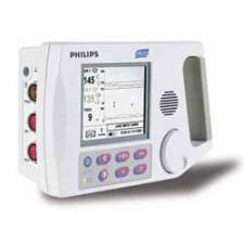
A standard echocardiogram is also known as a transthoracic echocardiogram (TTE), or cardiac ultrasound. In this case, the echocardiography transducer (or probe) is placed on the chest wall (or thorax) of the subject, and images are taken through the chest wall. This is a non-invasive, highly accurate and quick assessment of the overall health of the heart. A cardiologist can quickly assess a patient's heart valves and degree of heart muscle contraction (an indicator of the ejection fraction). The images are displayed on a monitor, and are recorded either by videotape (analog) or by digital techniques.
An echocardiogram can be used to evaluate all four chambers of the heart. It can determine strength of the heart, the condition of the heart valves, the lining of the heart (the pericardium), and the aorta. It can be used to detect a heart attack, enlargement or hypertrophy of the heart, infiltration of the heart with an abnormal substance. Weakness of the heart, cardiac tumors, and a variety of other findings can be diagnosed with an echocardiogram. With advanced measurements of the movement of the tissue with time (tissue doppler), it can measure diastolic function, fluid status, and dys-synchrony.
The TTE is highly accurate for identifying vegetations (masses consisting of a mixture of bacteria and blood clots), but the accuracy can be reduced in up to 20% of adults because of obesity, chronic obstructive pulmonary disease, chest-wall deformities, or otherwise technically difficult patients. TTE in adults is also of limited use for the structures at the back of the heart, such as the left atrial appendage. Transesophageal echocardiography may be more accurate than TTE because it excludes the variables previously mentioned and allows closer visualization of common sites for vegetations and other abnormalities. Transesophageal echocardiography also affords better visualization of prosthetic heart valves.
Transesophageal echocardiogram
This is an alternative way to perform an echocardiogram. A specialized probe containing an ultrasound transducer at its tip is passed into the patient's esophagus. This allows image and Doppler evaluation which can be recorded. This is known as a transesophageal echocardiogram, or TEE (TOE in the United Kingdom). The advantage of TEE over TTE is usually clearer images, especially of structures that are difficult to view transthoracicly (through the chest wall). The explanation for this is that the heart rests directly upon the esophagus leaving only millimeters that the ultrasound beam has to travel. This reduces the attenuation (weakening) of the ultrasound signal, generating a stronger return signal, ultimately enhancing image and Doppler quality. Comparatively, transthoracic ultrasound must first traverse skin, fat, ribs and lungs before reflecting off the heart and back to the probe before an image can be created. All these structures, along with the increased distance the beam must travel, weaken the ultrasound signal thus degrading the image and Doppler quality.
In adults, several structures can be evaluated and imaged better with the TEE, including the aorta, pulmonary artery, valves of the heart, both atria, atrial septum, left atrial appendage, and coronary arteries. TEE has a very high sensitivity for locating a blood clot inside the left atrium. While TTE can be performed quickly, easily and without pain to the patient, TEE requires a fasting patient, (the patient must not eat or drink after midnight on the day before the procedure), a team of medical personnel, takes longer to perform, is uncomfortable for the patient and has some risks associated with the procedure (esophageal perforation -- 1 in 10,000, and adverse reactions to the medication).
Before inserting the probe, conscious sedation is induced in the patient to ease the discomfort and to decrease the gag reflex, thus making the ultrasound probe easier to pass into the esophagus. Conscious sedation is a light sedation usually using the medications midazolam (a benzodiazepine with sedating, amnesiac qualities) and fentanyl. Sometimes a local anesthetic spray is used for the back of the throat, such a xylocaine and/or a jelly/lubricant anesthetic for the esophagus. Children are anesthetized. Unlike the TTE, the TEE is considered an invasive procedure.
How safe is echocardiography?
Echocardiography is extremely safe. There are no known risks from the clinical use of ultrasound during this type of testing.
How long does it take?
A brief examination in an uncomplicated case may be done within 15 to 20 minutes. The additional use of Doppler may add an additional 10 to 20 minutes. However, it may take up to an hour when there are multiple problems or when there are technical problems (for example, patients with lung disease, obesity, restlessness, and significant shortness of breath may be more difficult to image).
3-dimensional echocardiography
3D echocardiogram of a heart viewed from the apex3-D echocardiography is now possible, using an ultrasound probe with an array of transducers and an appropriate processing system. This enables detailed anatomical assessment of cardiac pathology, particularly valvular defects, and cardiomyopathies. The ability to slice the virtual heart in infinite planes in an anatomically appropriate manner and to reconstruct 3-dimensional images of anatomic structures make 3D echocardiography unique for the understanding of the congenitally malformed heart.
Sources:
http://www.asecho.org/i4a/pages/index.cfm?pageid=1
http://www.heartsite.com/html/echocardiogram.html
http://en.wikipedia.org/wiki/Echocardiography
http://images.google.com/images?gbv=2&hl=en&q=echocardiogram&sa=N&start=90&ndsp=18
Edited by: JD Sandham